Research
Cerebral Reorganization after Stroke
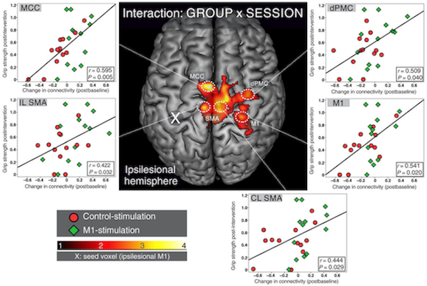 from Volz et al. 2016, Cerebral Cortex
The brain cannot regenerate neural tissue damaged by stroke. Improvement of functional impairment thus has to arise from repurposing intact neural circuitry to accommodate the neural processes necessary to compensate for the tissue loss. When patients are initial unable to move their hand (hemiplegia), different parts of the brain take over hand motor control to enable the patients to relearn to hand use. This process is referred to as cerebral reorganization. In our lab we study how the brain accomplishes successful reorganization using functional neuroimaging (fMRI, EEG) and non-invasive brain stimulation (TMS). Since functional reorganization mainly occurs in the first weeks after stroke – the so-called critical-period – we focus on the longitudinal assessment of acute stroke patients in a hospitalized setting. Our goal lies in the development of translational approaches to (i) predict the recovery-potential of individual patients and (ii) modulate reorganization to amplify motor recovery.
publications:
- Volz LJ, Vollmer M, Michely J, Fink GR, Grefkes C, Rothwell JC, Grefkes C (2017) Time-dependent functional role of the contralesional motor cortex after stroke. NeuroImage: Clinical: 16: 165-174
- Volz LJ, Rehme AK, Michely J, Nettekoven C, Eickhoff SB, Fink GR, Grefkes C (2016) Shaping Early Reorganization of Neural Networks Promotes Motor Function after Stroke. Cerebral Cortex 26(6):2882-2894
- Rehme AK, Volz LJ, Feis DL, Eickhoff SB, Fink GR, Grefkes C (2015) Early neuroimaging predictors of chronic motor disability after stroke. Human Brain Mapping 36(11):4553-65
- Rehme AK, Volz LJ, Feis DL, Bomilcar-Focke I, Liebig T, Eickhoff SB, Fink GR, Grefkes C (2015) Identifying neuroimaging markers of motor disability in acute stroke by machine learning techniques. Cerebral Cortex 25(9):3046-56
- Volz LJ, Sarfeld AS, Diekhoff S, Rehme AK, Pool EM, Eickhoff SB, Fink GR, Grefkes C (2015) Motor Cortex Excitability and Connectivity in chronic stroke – A multimodal model of functional reorganization. Brain Structure and Function 220(2):1093-107
- Volz LJ, Grefkes C (2013) Neurophysiological and Neuroimaging Predictors of Functional Recovery after Stroke. Klinische Neurophysiologie 44:238-246
Brain Stimulation
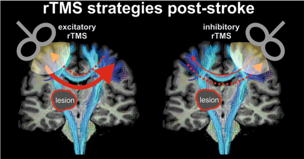 from Volz and Grefkes, 2015
Non-invasive brain stimulation such as transcranial magnetic stimulation (TMS) allows us to interact with the human brain in several ways. While single pulse TMS can be used to probe the excitability of motor system, repetitive TMS (rTMS) modulates neural activity of the stimulated circuitry beyond the duration of the stimulation. Finally, stimulation of cortical regions during task performance (online TMS) can help to investigate the functional role of cortical areas for the performed task in a causal way.
In our lab we investigate the mechanisms underlying plasticity-induction in the human brain and use online TMS to further our mechanistic understanding of regional contributions to complex and distributed brain functions such as motor control. Specifically, we aim at furthering our mechanistic understanding of cerebral reorganization and reward-dependent motor learning after stroke and ask the question how we can utilize rTMS to modulate post-stroke plasticity to benefit functional recovery.
publications:
- Volz LJ, Hamada M, Rothwell JC, Grefkes C (under review) What makes the muscle twitch more: a multimodal model of plasticity induction
- Krall SC, Volz LJ, Oberwelland E, Grefkes C, Fink GR, Konrad K (2016) The right temporoparietal junction in attention and social interaction: A transcranial magnetic stimulation study. Human Brain Mapping, 37(2), 796-807
- Volz LJ, Hamada M, Rothwell JC, Grefkes C (2015) What makes the muscle twitch: motor system connectivity and TMS-induced activity. Cerebral Cortex 25(9):2346-53
- Nettekoven C, Volz LJ, Leimbach M, Pool EM, Rehme AK, Eickhoff SB, Fink GR, Grefkes C (2015) Inter-individual variability in cortical excitability and motor network connectivity following multiple blocks of rTMS. NeuroImage 118: 209–218
- Nettekoven C, Volz LJ, Kutscha M, Pool EM, Rehme AK, Eickhoff SB, Fink GR, Grefkes C (2014) Dose- dependent effects of theta-burst rTMS on cortical excitability and resting-state connectivity of the human motor system. Journal of Neuroscience 34(20):6849-59
- Cárdenas-Morales L, Volz LJ, Rehme AK, Pool EM, Nettekoven C, Eickhoff SB, Fink GR, Grefkes C (2014) Network Connectivity and Individual Responses to Brain Stimulation in the Human Motor System. Cerebral Cortex 24(7):1697-707 (shared first-authorship)
- Volz LJ, Benali A, Mix A, Neubacher U, Funke K (2013) Dose-dependence of changes in cortical protein expression induced with repeated transcranial magnetic theta-burst stimulation in the rat. Brain Stimulation 6(4):598-606
Brain Network Connectivity
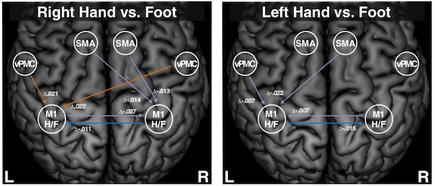 from Volz et al. 2015, NeuroImage
Human behavior is typically not controlled by single brain regions but results from the fine-tuned interaction of multiple brain areas. Analyzing the connectivity of remote but inter-connected brain areas can help to understand the neural mechanisms underlying human behavior and its impairment due to diseases.
We use a multimodal approach of structural and functional MRI to further our insights into the functional anatomy of how the brain integrates information to enable the human condition. Specifically, we aim at integrating insights from a multimodal perspective to understand how network structure predetermines function with an emphasis on network alterations due to stroke lesions or degenerative processes.
publications:
- Greene C, Cieslak M, Volz LJ, Hensel L, Grefkes C, Rose K, Grafton ST (2019) Finding maximally disconnected subnetworks with shortest path tractography. NeuroImage: Clinical 18: doi.org/10.1016/j.nicl.2019.101903
- Cieslak M, Meiring W, Brennan T, Greene C, Volz LJ, Vettel JM, Suri S, Grafton ST (2018) Compositional measures of diffusion anisotropy and asymmetry. Biomedical Imaging (ISBI 2018), IEEE 15th International Symposium: 123-126
- Volz LJ, Cieslak M, Grafton ST (2018) Compartmentalizing white matter reduces profound variability of anisotropy measures. Brain Structure and Function 223(2), 635-651
- Cieslak M, Brennan T, Meiring W, Volz LJ, Asturias A, Surib S, Grafton ST (2018) Analytic tractography: A closed-form solution for estimating local white matter connectivity with diffusion MRI. NeuroImage 169: 473–484
- Michely J, Volz LJ, Hoffstaedter F, Eickhoff SB, Fink GR, Grefkes C (2018) Network connectivity of motor control in the ageing brain. NeuroImage: Clinical 18: 443-455
- Volz LJ, Eickhoff SB, Pool EM, Fink GR, Grefkes C (2015) Differential Modulation of Motor Network Connectivity during Movements of the Upper and Lower Limbs. NeuroImage 119: 44–53
- Michely J, Volz LJ, Barbe M, Hoffstaedter F, Viswanathan S, Timmermann L, Eickhoff SB, Fink GR, Grefkes C (2015) Dopaminergic modulation of motor network dynamics in Parkinson’s disease. Brain 138(Pt 3):664-78
Decision-making and Learning
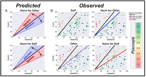 from Volz et al. 2017, PNAS
Decision-making has a profound influence on our daily life and personal development. Implementing new control-policies which allow to change our decision-making behavior and skill-acquisition through learning enable us to adapt to our ever changing environment. Understanding the neural basis of decision-making and learning thus is paramount not only from a theoretical perspective, but has many practical implications ranging from understanding human behavior in moral or social situations to how we compensate brain lesions by adapting motor control-policies and learning new motor skills to compensate functional impairment. In particular, the question how cerebral reorganization after stroke impacts on and interacts with decision-making and skill learning (e.g. in the motor domain) remains poorly understood. Our goal is to combine increasingly complex behavioral readouts reflecting motor learning and changes in decision-making patterns with multimodal neural data in stroke patients to elucidate how the heightened level of neural plasticity in the critical-period early after stroke is shaped by and allows motor-skill learning and formation of new decision-making policies. Specifically, we aim at increasing our mechanistic understanding of how changes in learning and decision-making facilitate functional recovery after brain lesions and developing novel treatment approaches by modulating such mechanisms, e.g. via suitable reinforcement strategies (reward/punishment), neuropharmacological interventions or non-invasive brain stimulation.
publications:
- Volz LJ et al. (in preperation)
- Layher E, Santander T, Volz LJ, Miller MB (2018) Failure to affect decision criteria during recognition memory with continuous theta burst stimulation. Frontiers in Neuroscience 12: 705
- Volz LJ, Welborn LB, Gobel MS, Gazzaniga MS, Grafton ST (2017) Harm to self outweighs benefit to others in moral decision making. Proceedings of the Nation Academy of Sciences 114 (30): 7963-7968
Split-brain and Consciousness
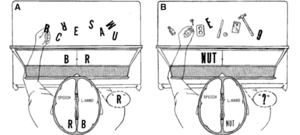 from Volz and Gazzaniga 2017, Brain
The insights gained from testing these so called ‘split-brain’ patients, which underwent sectioning of the corpus callosum as a treatment for severe epilepsy, have profoundly shaped the field of cognitive neuroscience and helped to establish seminal information processing models for how the brain governs behavior and cognition. For example, split-brain research has highlighted distinct interpretive capacities of both cerebral hemispheres and contributed important pieces to the puzzle of deciphering the neurobiological foundations that give rise to our conscious experience of the world.
publications:
- Volz LJ, Hillyard SA, Miller MB, Gazzaniga MS (2018) Unifying control over the body: consciousness and cross- cueing in split-brain patients. Brain 141 (3), e15
- Volz LJ, Gazzaniga MS (2017) Interaction in isolation - 50 years of insights from split-brain research. Brain 140 (7): 2051-2060
Translational Neuroimaging
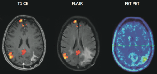 from Volz et al. 2018, Q J Nucl Med Mol Imaging
Advanced functional and structural neuroimaging methods such as diffusion MRI or fMRI offer the potential to further our mechanistic understanding of psychiatric and neurological disease and may help to create new diagnostic tools enabling earlier and more accurate diagnosis and even prognosis on the level of individual patients. Our goal is to advance the clinical utility of neuroimaging by applying novel methods to patient populations in cooperation with both methodological experts and specialized clinicians.
publications:
- Cohrs G, Huhndorf M, Niemczyk N, Volz LJ, Bernsmeier A, Singhal A, Larsen N, Synowitz M, Knerlich- Lukoschus F (2018) MRI in mild pediatric traumatic brain injury: diagnostic overkill or useful tool? Child’s nervous system 34 (7), 1345-1352
- Volz LJ, Kocher M, Lohmann P, Shah NJ, Fink GR, Galldiks N (2018) Functional magnetic resonance imaging in glioma patients: from clinical applications to future perspectives. The quarterly journal of nuclear medicine and molecular imaging 62(3):295-302
- Pedrosa DJ, Nelles C, Brown P, Volz LJ, Pelzer EA, Tittgemeyer M, Brittain JS, Timmermann L (2017) The differentiated networks related to Essential tremor onset and its amplitude modulation after alcohol intake. Experimental Neurology 297: 50–61
